CT image visual quantitative evaluation and clinical classification of coronavirus disease (COVID-19).
Authors of this article are:
Li K, Fang Y, Li W, Pan C, Qin P, Zhong Y, Liu X, Huang M, Liao Y, Li S.
A summary of the article is shown below:
OBJECTIVES: To explore the relationship between the imaging manifestations and clinical classification of COVID-19.METHODS: We conducted a retrospective single-center study on patients with COVID-19 from Jan. 18, 2020 to Feb. 7, 2020 in Zhuhai, China. Patients were divided into 3 types based on Chinese guideline: mild (patients with minimal symptoms and negative CT findings), common, and severe-critical (patients with positive CT findings and different extent of clinical manifestations). CT visual quantitative evaluation was based on summing up the acute lung inflammatory lesions involving each lobe, which was scored as 0 (0%), 1 (1-25%), 2 (26-50%), 3 (51-75%), or 4 (76-100%), respectively. The total severity score (TSS) was reached by summing the five lobe scores. The consistency of two observers was evaluated. The TSS was compared with the clinical classification. ROC was used to test the diagnosis ability of TSS for severe-critical type.RESULTS: This study included 78 patients, 38 males and 40 females. There were 24 mild (30.8%), 46 common (59.0%), and 8 severe-critical (10.2%) cases, respectively. The median TSS of severe-critical-type group was significantly higher than common type (p < 0.001). The ICC value of the two observers was 0.976 (95% CI 0.962-0.985). ROC analysis showed the area under the curve (AUC) of TSS for diagnosing severe-critical type was 0.918. The TSS cutoff of 7.5 had 82.6% sensitivity and 100% specificity.CONCLUSIONS: The proportion of clinical mild-type patients with COVID-19 was relatively high; CT was not suitable for independent screening tool. The CT visual quantitative analysis has high consistency and can reflect the clinical classification of COVID-19.KEY POINTS: • CT visual quantitative evaluation has high consistency (ICC value of 0.976) among the observers. The median TSS of severe-critical type group was significantly higher than common type (p < 0.001). • ROC analysis showed the area under the curve (AUC) of TSS for diagnosing severe-critical type was 0.918 (95% CI 0.843-0.994). The TSS cutoff of 7.5 had 82.6% sensitivity and 100% specificity. • The proportion of confirmed COVID-19 patients with normal chest CT was relatively high (30.8%); CT was not a suitable screening modality.
Check out the article’s website on Pubmed for more information:
[link-preview url=https://www.ncbi.nlm.nih.gov/pubmed/32215691 forceshot=true]
This article is a good source of information and a good way to become familiar with topics such as: COVID-19; Classification; Quantitative evaluation; Tomography, X-ray computed.
New Research Kits from MOLECULAR DEPOT
-
- Sale!
- Interference, Research Kits
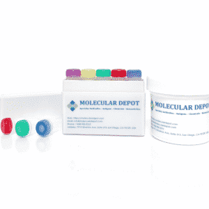
Interference Test Kit
- Original price was: $995.00.$495.00Current price is: $495.00.
-
- Sale!
- Research Kits
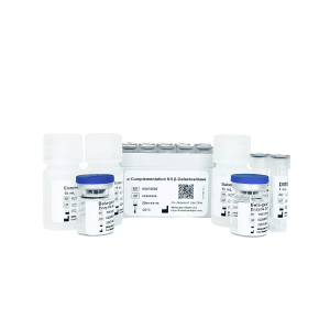
Alpha Complementation Kit Beta-galactosidase
- Original price was: $1,795.00.$895.00Current price is: $895.00.
-
- Sale!
- Research Kits
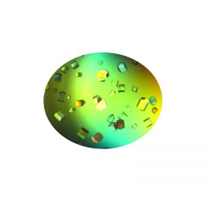
Crystallization Screen for Protein-DNA Complexes
- Original price was: $1,995.00.$995.00Current price is: $995.00.
-
- Sale!
- Molecular Biology, Research Kits
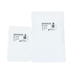
NativeFolder Bacterial Culture Medium
- Original price was: $795.00.$295.00Current price is: $295.00.
-
- Sale!
- Research Kits
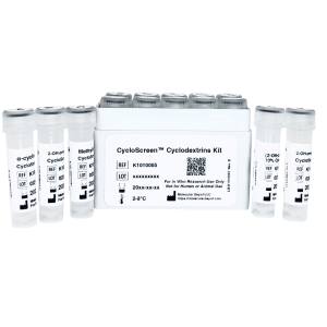
CycloScreen Cyclodextrins Kit
- Original price was: $695.00.$350.00Current price is: $350.00.
-
- Sale!
- Research Kits
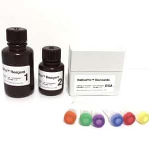
NativePro Bradford Protein Assay Kit (Automated)
- Original price was: $995.00.$495.00Current price is: $495.00.
-
- Sale!
- Interference, Research Kits
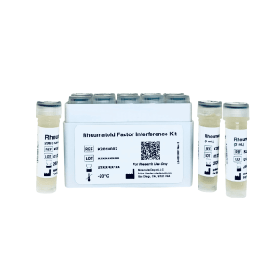
Rheumatoid Factor Interference Kit
- Original price was: $1,495.00.$695.00Current price is: $695.00.
-
- Sale!
- Interference, Research Kits
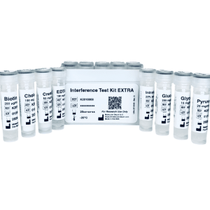
Interference Test Kit EXTRA
- Original price was: $1,295.00.$695.00Current price is: $695.00.
-
- Sale!
- Interference, Research Kits
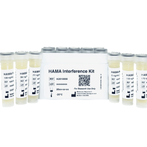
HAMA Interference Kit
- Original price was: $1,295.00.$695.00Current price is: $695.00.
-
- Sale!
- Buffers & Solutions, ELISA, Research Kits
ELISA Microplate Production Kit
- Original price was: $795.00.$495.00Current price is: $495.00.






New Antibodies from MOLECULAR DEPOT
-
- Sale!
- Antibodies
Zebrafish wnt8b Antibody (Center)
- Original price was: $1,295.00.$695.00Current price is: $695.00.
-
- Sale!
- Antibodies
TRIB2 Antibody (N-Term)
- Original price was: $1,295.00.$695.00Current price is: $695.00.
-
- Sale!
- Antibodies
NTN1 Antibody (C-Term)
- Original price was: $1,295.00.$695.00Current price is: $695.00.
-
- Sale!
- Antibodies
NME1 Antibody
- Original price was: $1,295.00.$695.00Current price is: $695.00.
-
- Sale!
- Antibodies
Parp9 Antibody (C-Term)
- Original price was: $1,295.00.$695.00Current price is: $695.00.
-
- Sale!
- Antibodies
CDK1 Antibody
- Original price was: $1,295.00.$695.00Current price is: $695.00.
-
- Sale!
- Antibodies
DANRE hoxc11a Antibody (N-Term)
- Original price was: $1,295.00.$695.00Current price is: $695.00.
-
- Sale!
- Antibodies
VDR Antibody (Center)
- Original price was: $1,295.00.$695.00Current price is: $695.00.
-
- Sale!
- Antibodies
ITIH4 Antibody (C-Term)
- Original price was: $1,295.00.$695.00Current price is: $695.00.
-
- Sale!
- Antibodies
SEMA4B Antibody (C-Term)
- Original price was: $1,295.00.$695.00Current price is: $695.00.

New Proteins from MOLECULAR DEPOT
-
- Sale!
- Conjugates, Proteins
Captopril HSA Biotin Conjugate
- Original price was: $1,995.00.$995.00Current price is: $995.00.
-
- Sale!
- Proteins
PKC-α, Active, GST-tagged from Xanopus sp.
- Original price was: $1,595.00.$795.00Current price is: $795.00.
-
- Sale!
- Conjugates, Proteins
Prilocain BSA Biotin Conjugate
- Original price was: $1,995.00.$995.00Current price is: $995.00.
-
- Sale!
- Conjugates, Proteins
T3 (Triiodothyronine) BSA Biotin Conjugate
- Original price was: $1,995.00.$995.00Current price is: $995.00.
-
- Sale!
- Conjugates, Proteins
Pipemidic Acid BSA Biotin Conjugate
- Original price was: $1,995.00.$995.00Current price is: $995.00.
-
- Sale!
- Proteins
PKD2 Protein, Active (Recombinant Human)
- Original price was: $1,795.00.$895.00Current price is: $895.00.
-
- Sale!
- Conjugates, Proteins
Meropenem BSA Biotin Conjugate
- Original price was: $1,995.00.$995.00Current price is: $995.00.
-
- Sale!
- Conjugates, Proteins
Piperacillin BSA Biotin Conjugate
- Original price was: $1,995.00.$995.00Current price is: $995.00.
-
- Sale!
- Proteins
Protein A Peroxidase from Staphylococcus aureus/horseradish
- Original price was: $1,195.00.$595.00Current price is: $595.00.
-
- Sale!
- Conjugates, Proteins
Tobramycin BSA Biotin Conjugate
- Original price was: $1,995.00.$995.00Current price is: $995.00.

New Chemicals from MOLECULAR DEPOT
-
- Sale!
- Chemicals, Interference, Lipids
Triglyceride Mix for Interference Testing
- Original price was: $895.00.$395.00Current price is: $395.00.
-
- Sale!
- Chemicals
Microparticle Stabilizer Solution
- Price range: $295.00 through $495.00
-
- Sale!
- Buffers & Solutions, Chemicals
Enzyme Acceptor Stabilization Buffer
- Original price was: $495.00.$250.00Current price is: $250.00.
-
- Sale!
- Buffers & Solutions, Chemicals
Enzyme Donor Stabilization Buffer
- Original price was: $495.00.$250.00Current price is: $250.00.
-
- Sale!
- Chemicals, Conjugates
Progesterone Biotin Conjugate Solution
- Original price was: $995.00.$595.00Current price is: $595.00.
-
- Sale!
- Buffers & Solutions, Chemicals
Microparticle Activation Buffer
- Price range: $395.00 through $695.00
-
- Sale!
- Buffers & Solutions, Chemicals
Microparticle Washing Buffer
- Price range: $395.00 through $695.00
-
- Sale!
- Buffers & Solutions, Chemicals
Microparticle Blocking Buffer
- Price range: $395.00 through $695.00
-
- Sale!
- Buffers & Solutions, Chemicals
Latex Microparticles Anti Aggregation Agent
- Price range: $295.00 through $3,500.00
-
- Sale!
- Chemicals
CPRG (Chlorophenol red-β-D-galactopyranoside)
- Original price was: $850.00.$395.00Current price is: $395.00.