
Radiology-pathology correlation in staging of liver fibrosis using superb microvascular imaging.
Authors of this article are:
Balık AÖ, Kılıçoğlu ZG, Görmez A, Özkara S.
A summary of the article is shown below:
PURPOSE: Progression of liver fibrosis to end-stage disease can potentially be prevented with antiviral treatment. Thus, diagnosis of fibrosis is important in determining treatment protocols. This study aims first, to determine the sensitivity of a novel Doppler method, superb microvascular imaging (SMI), in comparison to other Doppler methods, in detecting small vascular structures of the liver; and second, to choose the best method among these Doppler applications to determine the morphological changes that occur due to chronic fibrosis. By doing so, the study would be able to provide an ultrasound grading that might differentiate and predict mild and severe liver fibrosis, thus giving rise to a possible alternative to biopsy.METHODS: 43 patients diagnosed with chronic hepatitis and scheduled for liver biopsy were included. Color Doppler, Power Doppler, ADF (advanced dynamic flow) Doppler, cSMI (color SMI) and mSMI (monochrome SMI) Doppler was performed in subcapsular areas of right anterior lobe. Depth from the capsule of the most peripherally located detectable vessel was measured for each Doppler subgroup. ANOVA test was applied after than Levene’s test was used to homogeneity of variance and Student-Newman-Keuls test was applied as post-hoc tests. Appearance of the vascular tree was categorized into four groups, and correlated with pathology results. ROC curve analysis was used to determine if this Doppler classification was statistically significant in differentiating mild and severe forms of fibrosis. Finally, multiple regression analysis determined which of the Doppler parameter was statistically significant in predicting severity.RESULTS: mSMI and cSMI were found to be superior to other Doppler techniques in detecting the most superficially located vessels of the liver; 4.4 mm and 3.3 mm deep from the capsule, respectively (P < 0.001). Among the changes identified in the vascular tree, small vessel blunting was the most prevalent finding in predicting the presence of severe fibrosis; multiple regression test, (t=5.969, P < 0.0001). ROC analysis identified that the presence of at least two pathological findings in the vascular tree was highly predictive of severe fibrosis; AUC= 0.881, sensitivity 86.67%, specificity 89.29%, positive and negative predictive values 8.09 and 0.15, respectively.CONCLUSION: Our study proves that SMI is superior to other Doppler techniques in detected the smallest vessels visible to ultrasound. Using this method, it is possible to determine the vascular changes in terms of blunting and tortuosity and thus predict the severity of fibrosis. This method might be a practical alternative to biopsy.
Check out the article’s website on Pubmed for more information:
[link-preview url=https://www.ncbi.nlm.nih.gov/pubmed/31287429 forceshot=true]
This article is a good source of information and a good way to become familiar with topics such as: n/a.
New Chemicals from MOLECULAR DEPOT
-
- Sale!
- Chemicals, Interference, Lipids
Triglyceride Mix for Interference Testing
- Original price was: $895.00.$395.00Current price is: $395.00.
-
- Sale!
- Chemicals
Microparticle Stabilizer Solution
- Price range: $295.00 through $495.00
-
- Sale!
- Buffers & Solutions, Chemicals
Enzyme Acceptor Stabilization Buffer
- Original price was: $495.00.$250.00Current price is: $250.00.
-
- Sale!
- Buffers & Solutions, Chemicals
Enzyme Donor Stabilization Buffer
- Original price was: $495.00.$250.00Current price is: $250.00.
-
- Sale!
- Chemicals, Conjugates
Progesterone Biotin Conjugate Solution
- Original price was: $995.00.$595.00Current price is: $595.00.
-
- Sale!
- Buffers & Solutions, Chemicals
Microparticle Activation Buffer
- Price range: $395.00 through $695.00
-
- Sale!
- Buffers & Solutions, Chemicals
Microparticle Washing Buffer
- Price range: $395.00 through $695.00
-
- Sale!
- Buffers & Solutions, Chemicals
Microparticle Blocking Buffer
- Price range: $395.00 through $695.00
-
- Sale!
- Buffers & Solutions, Chemicals
Latex Microparticles Anti Aggregation Agent
- Price range: $295.00 through $3,500.00
-
- Sale!
- Chemicals
CPRG (Chlorophenol red-β-D-galactopyranoside)
- Original price was: $850.00.$395.00Current price is: $395.00.






New Proteins from MOLECULAR DEPOT
-
- Sale!
- Conjugates, Proteins
Captopril HSA Biotin Conjugate
- Original price was: $1,995.00.$995.00Current price is: $995.00.
-
- Sale!
- Proteins
PKC-α, Active, GST-tagged from Xanopus sp.
- Original price was: $1,595.00.$795.00Current price is: $795.00.
-
- Sale!
- Conjugates, Proteins
Prilocain BSA Biotin Conjugate
- Original price was: $1,995.00.$995.00Current price is: $995.00.
-
- Sale!
- Conjugates, Proteins
T3 (Triiodothyronine) BSA Biotin Conjugate
- Original price was: $1,995.00.$995.00Current price is: $995.00.
-
- Sale!
- Conjugates, Proteins
Pipemidic Acid BSA Biotin Conjugate
- Original price was: $1,995.00.$995.00Current price is: $995.00.
-
- Sale!
- Proteins
PKD2 Protein, Active (Recombinant Human)
- Original price was: $1,795.00.$895.00Current price is: $895.00.
-
- Sale!
- Conjugates, Proteins
Meropenem BSA Biotin Conjugate
- Original price was: $1,995.00.$995.00Current price is: $995.00.
-
- Sale!
- Conjugates, Proteins
Piperacillin BSA Biotin Conjugate
- Original price was: $1,995.00.$995.00Current price is: $995.00.
-
- Sale!
- Proteins
Protein A Peroxidase from Staphylococcus aureus/horseradish
- Original price was: $1,195.00.$595.00Current price is: $595.00.
-
- Sale!
- Conjugates, Proteins
Tobramycin BSA Biotin Conjugate
- Original price was: $1,995.00.$995.00Current price is: $995.00.

New Antibodies from MOLECULAR DEPOT
-
- Sale!
- Antibodies
SUMO1 Antibody (C-Term)
- Original price was: $1,295.00.$695.00Current price is: $695.00.
-
- Sale!
- Antibodies
IL18RAP Antibody (N-Term)
- Original price was: $1,295.00.$695.00Current price is: $695.00.
-
- Sale!
- Antibodies
DENR Antibody
- Original price was: $1,295.00.$695.00Current price is: $695.00.
-
- Sale!
- Antibodies
Mouse DHFR Antibody (C-Term)
- Original price was: $1,295.00.$695.00Current price is: $695.00.
-
- Sale!
- Antibodies
BCL6B Antibody (Center)
- Original price was: $1,295.00.$695.00Current price is: $695.00.
-
- Sale!
- Antibodies
RSK4 Antibody (N-Term)
- Original price was: $1,295.00.$695.00Current price is: $695.00.
-
- Sale!
- Antibodies
IRF3 Antibody
- Original price was: $1,295.00.$695.00Current price is: $695.00.
-
- Sale!
- Antibodies
DAAM1-T361 Antibody (N-Term)
- Original price was: $1,295.00.$695.00Current price is: $695.00.
-
- Sale!
- Antibodies
RHEX Antibody (C-Term)
- Original price was: $1,295.00.$695.00Current price is: $695.00.
-
- Sale!
- Antibodies
Myoglobin Antibody
- Original price was: $1,295.00.$695.00Current price is: $695.00.
New Research Kits from MOLECULAR DEPOT
-
- Sale!
- Interference, Research Kits
Interference Test Kit
- Original price was: $995.00.$495.00Current price is: $495.00.
-
- Sale!
- Research Kits
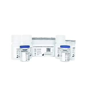
Alpha Complementation Kit Beta-galactosidase
- Original price was: $1,795.00.$895.00Current price is: $895.00.
-
- Sale!
- Research Kits
Crystallization Screen for Protein-DNA Complexes
- Original price was: $1,995.00.$995.00Current price is: $995.00.
-
- Sale!
- Molecular Biology, Research Kits
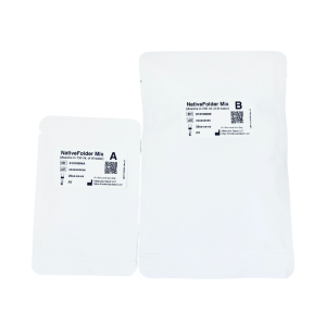
NativeFolder Bacterial Culture Medium
- Original price was: $795.00.$295.00Current price is: $295.00.
-
- Sale!
- Research Kits
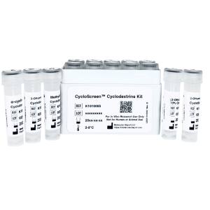
CycloScreen Cyclodextrins Kit
- Original price was: $695.00.$350.00Current price is: $350.00.
-
- Sale!
- Research Kits
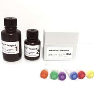
NativePro Bradford Protein Assay Kit (Automated)
- Original price was: $995.00.$495.00Current price is: $495.00.
-
- Sale!
- Interference, Research Kits
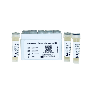
Rheumatoid Factor Interference Kit
- Original price was: $1,495.00.$695.00Current price is: $695.00.
-
- Sale!
- Interference, Research Kits
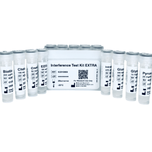
Interference Test Kit EXTRA
- Original price was: $1,295.00.$695.00Current price is: $695.00.
-
- Sale!
- Interference, Research Kits
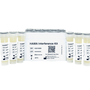
HAMA Interference Kit
- Original price was: $1,295.00.$695.00Current price is: $695.00.
-
- Sale!
- Buffers & Solutions, ELISA, Research Kits
ELISA Microplate Production Kit
- Original price was: $795.00.$495.00Current price is: $495.00.