
Microscope and micro-camera assessment of Schneiderian membrane perforation via transcrestal sinus floor elevation: A randomized ex vivo study.
Authors of this article are:
Gargallo-Albiol J, Sinjab KH, Barootchi S, Chan HL, Wang HL.
A summary of the article is shown below:
OBJECTIVE: We sought to assess the effectiveness of using a microscope and non-invasive camera for assessing sinus membrane perforations during transcrestal sinus floor elevation (TSFE).MATERIALS AND METHODS: Five fresh human cadaver heads corresponding to eight maxillary sinuses (six bilateral and two unilateral) underwent 4 TSFEs per sinus (a total of 32 single site elevations). Each elevation was randomly assigned to receive a three or six mm membrane elevation height (MEH). A microscope and micro-camera were used to assess the sinus membrane perforation. Afterwards, radiological and clinical membrane perforation assessments were performed. The statistical analysis results are expressed using the means, standard deviations, range values of the residual ridge height (RRH), residual ridge width (RRW), sinus membrane thickness (SMT) and incidence of perforation (IoP). Generalized linear methods were used to test for the correlation of RRH and MEH to the microscope and micro-camera perforation assessments and the correlation of microscope and micro-camera assessments with the post-operative CBCT and crestal liquid evaluation.RESULTS: The cumulative percentage of IoP was 40.62%, (23.07% with 3 mm MEH, and 76.92% with 6 mm MEH, p < 0.05). The perforation assessed using either the microscope or micro-camera coincided with the post-operative CBCT and crestal liquid assessment in 87.55% sites. No significant correlation was found between the microscope or micro-camera assessments with RRH or MEH.CONCLUSION: Application of a microscope and micro-camera during transcrestal sinus floor elevation may allow the detection of the integrity of the Schneiderian membrane with greater than 85% accuracy in this ex vivo model.© 2019 John Wiley & Sons A/S. Published by John Wiley & Sons Ltd.
Check out the article’s website on Pubmed for more information:
[link-preview url=https://www.ncbi.nlm.nih.gov/pubmed/31059148 forceshot=true]
This article is a good source of information and a good way to become familiar with topics such as: clinical assessment;diagnosis;sinus floor elevation.
New Chemicals from MOLECULAR DEPOT
-
- Sale!
- Chemicals, Interference, Lipids
Triglyceride Mix for Interference Testing
- Original price was: $895.00.$395.00Current price is: $395.00.
-
- Sale!
- Chemicals
Microparticle Stabilizer Solution
- Price range: $295.00 through $495.00
-
- Sale!
- Buffers & Solutions, Chemicals
Enzyme Acceptor Stabilization Buffer
- Original price was: $495.00.$250.00Current price is: $250.00.
-
- Sale!
- Buffers & Solutions, Chemicals
Enzyme Donor Stabilization Buffer
- Original price was: $495.00.$250.00Current price is: $250.00.
-
- Sale!
- Chemicals, Conjugates
Progesterone Biotin Conjugate Solution
- Original price was: $995.00.$595.00Current price is: $595.00.
-
- Sale!
- Buffers & Solutions, Chemicals
Microparticle Activation Buffer
- Price range: $395.00 through $695.00
-
- Sale!
- Buffers & Solutions, Chemicals
Microparticle Washing Buffer
- Price range: $395.00 through $695.00
-
- Sale!
- Buffers & Solutions, Chemicals
Microparticle Blocking Buffer
- Price range: $395.00 through $695.00
-
- Sale!
- Buffers & Solutions, Chemicals
Latex Microparticles Anti Aggregation Agent
- Price range: $295.00 through $3,500.00
-
- Sale!
- Chemicals
CPRG (Chlorophenol red-β-D-galactopyranoside)
- Original price was: $850.00.$395.00Current price is: $395.00.






New Proteins from MOLECULAR DEPOT
-
- Sale!
- Conjugates, Proteins
Captopril HSA Biotin Conjugate
- Original price was: $1,995.00.$995.00Current price is: $995.00.
-
- Sale!
- Proteins
PKC-α, Active, GST-tagged from Xanopus sp.
- Original price was: $1,595.00.$795.00Current price is: $795.00.
-
- Sale!
- Conjugates, Proteins
Prilocain BSA Biotin Conjugate
- Original price was: $1,995.00.$995.00Current price is: $995.00.
-
- Sale!
- Conjugates, Proteins
T3 (Triiodothyronine) BSA Biotin Conjugate
- Original price was: $1,995.00.$995.00Current price is: $995.00.
-
- Sale!
- Conjugates, Proteins
Pipemidic Acid BSA Biotin Conjugate
- Original price was: $1,995.00.$995.00Current price is: $995.00.
-
- Sale!
- Proteins
PKD2 Protein, Active (Recombinant Human)
- Original price was: $1,795.00.$895.00Current price is: $895.00.
-
- Sale!
- Conjugates, Proteins
Meropenem BSA Biotin Conjugate
- Original price was: $1,995.00.$995.00Current price is: $995.00.
-
- Sale!
- Conjugates, Proteins
Piperacillin BSA Biotin Conjugate
- Original price was: $1,995.00.$995.00Current price is: $995.00.
-
- Sale!
- Proteins
Protein A Peroxidase from Staphylococcus aureus/horseradish
- Original price was: $1,195.00.$595.00Current price is: $595.00.
-
- Sale!
- Conjugates, Proteins
Tobramycin BSA Biotin Conjugate
- Original price was: $1,995.00.$995.00Current price is: $995.00.

New Antibodies from MOLECULAR DEPOT
-
- Sale!
- Antibodies
SUMO1 Antibody (C-Term)
- Original price was: $1,295.00.$695.00Current price is: $695.00.
-
- Sale!
- Antibodies
IL18RAP Antibody (N-Term)
- Original price was: $1,295.00.$695.00Current price is: $695.00.
-
- Sale!
- Antibodies
DENR Antibody
- Original price was: $1,295.00.$695.00Current price is: $695.00.
-
- Sale!
- Antibodies
Mouse DHFR Antibody (C-Term)
- Original price was: $1,295.00.$695.00Current price is: $695.00.
-
- Sale!
- Antibodies
BCL6B Antibody (Center)
- Original price was: $1,295.00.$695.00Current price is: $695.00.
-
- Sale!
- Antibodies
RSK4 Antibody (N-Term)
- Original price was: $1,295.00.$695.00Current price is: $695.00.
-
- Sale!
- Antibodies
IRF3 Antibody
- Original price was: $1,295.00.$695.00Current price is: $695.00.
-
- Sale!
- Antibodies
DAAM1-T361 Antibody (N-Term)
- Original price was: $1,295.00.$695.00Current price is: $695.00.
-
- Sale!
- Antibodies
RHEX Antibody (C-Term)
- Original price was: $1,295.00.$695.00Current price is: $695.00.
-
- Sale!
- Antibodies
Myoglobin Antibody
- Original price was: $1,295.00.$695.00Current price is: $695.00.
New Research Kits from MOLECULAR DEPOT
-
- Sale!
- Interference, Research Kits
Interference Test Kit
- Original price was: $995.00.$495.00Current price is: $495.00.
-
- Sale!
- Research Kits
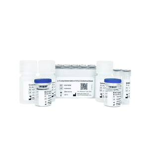
Alpha Complementation Kit Beta-galactosidase
- Original price was: $1,795.00.$895.00Current price is: $895.00.
-
- Sale!
- Research Kits
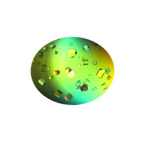
Crystallization Screen for Protein-DNA Complexes
- Original price was: $1,995.00.$995.00Current price is: $995.00.
-
- Sale!
- Molecular Biology, Research Kits
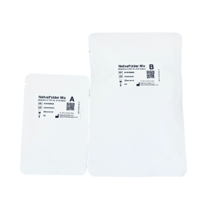
NativeFolder Bacterial Culture Medium
- Original price was: $795.00.$295.00Current price is: $295.00.
-
- Sale!
- Research Kits
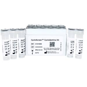
CycloScreen Cyclodextrins Kit
- Original price was: $695.00.$350.00Current price is: $350.00.
-
- Sale!
- Research Kits
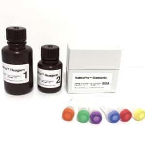
NativePro Bradford Protein Assay Kit (Automated)
- Original price was: $995.00.$495.00Current price is: $495.00.
-
- Sale!
- Interference, Research Kits
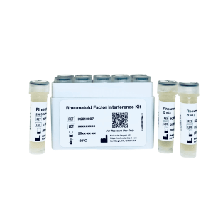
Rheumatoid Factor Interference Kit
- Original price was: $1,495.00.$695.00Current price is: $695.00.
-
- Sale!
- Interference, Research Kits
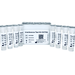
Interference Test Kit EXTRA
- Original price was: $1,295.00.$695.00Current price is: $695.00.
-
- Sale!
- Interference, Research Kits
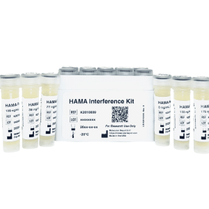
HAMA Interference Kit
- Original price was: $1,295.00.$695.00Current price is: $695.00.
-
- Sale!
- Buffers & Solutions, ELISA, Research Kits
ELISA Microplate Production Kit
- Original price was: $795.00.$495.00Current price is: $495.00.