
Internal Mammary Lymph Node Visualization as a Sentinel Sonographic Sign of Tuberculous Pleurisy.
Authors of this article are:
Messineo L, Quadri F, Valsecchi A, Lonni S, Palmiotti A, Giampietro M.
A summary of the article is shown below:
in English, GermanHINTERGRUND: Die tuberkulöse Pleuritis ist eine der primären Stellen der extrapulmonalen Tuberkulose. Derzeit fehlt es den Ärzten jedoch an diagnostischen Mitteln für die Früherkennung, wenn typische Anzeichen und Symptome fehlen. Mit dieser Studie wollten wir den Zusammenhang zwischen interner Mamma-Adenopathie und tuberkulöser Pleuritis (TP) untersuchen.METHODEN: 60 Patienten mit der post-thorakoskopischen histologischen Diagnose einer Granulomatose oder akuten infektiösen Pleuritis wurden retrospektiv aufgenommen. Bei allen wurde eine Thorax-Sonografie durchgeführt und/oder es lagen CT-Aufnahmen vor. Mindestens 2 auf Thorax-Sonografie spezialisierte Ärzte reanalysierten die sonografischen Aufnahmen im Hinblick auf interne Mamma-Adenopathien. Entsprechende Befunde wurden mit den CT-Daten verglichen.ERGEBNISSE: Die Thorax-Sonografie zeigte eine interne Mamma-Adenopathie ipsilateral zum Pleuraerguss bei 97 % der 29 Patienten mit diagnostizierter TP und bei 13 % der Patienten, die eine akute infektiöse Pleuritis hatten (p < 0,001). Die ROC-Analyse („receiver operator characteristic“) ergab für diese Methode eine Sensitivität von 97 % und eine Spezifität von 87 % für die Vorhersage einer TP („area under the curve“ 0,92 ± 0,04, p < 0,001). Die CT-Detektionsleistung und die Knotenmessungen waren signifikant ähnlich (p < 0,001).SCHLUSSFOLGERUNG: Die sonografische Darstellung der inneren Brustknoten ipsilateral zum Pleuraerguss kann zu einem Sentinelmarker für TP werden, der zur Frühdiagnose beiträgt oder bei fraglichen Fällen das diagnostische Management in Richtung invasiver Verfahren lenkt.BACKGROUND: Tuberculous pleurisy is one of the primary sites of extrapulmonary tuberculosis, but clinicians currently lack the diagnostic tools necessary for early recognition in the absence of typical signs and symptoms. With this study, we aimed to test the association between internal mammary adenopathies and tuberculous pleurisy (TP).METHODS: 60 patients with a post-thoracoscopic histological diagnosis of granulomatosis or acute infective pleurisy were retrospectively enrolled. All of them had chest sonography and/or CT scan data available. At least two expert chest sonography physicians re-analyzed the sonography images to look for any internal mammary adenopathy. Such findings were compared to the CT data.RESULTS: Chest sonography showed internal mammary adenopathy ipsilateral to the pleural effusion in 97 % of 29 patients who had a diagnosis of TP, and in 13 % of those with an acute infective pleurisy (p < 0.001). Receiver operator characteristic analysis revealed 97 % sensitivity and 87 % specificity for this technique in predicting TP (area under curve 0.92 ± 0.04, p < 0.001). CT detection power and node measures were significantly similar (p < 0.001).CONCLUSION: Sonographic internal mammary node visualization ipsilateral to the pleural effusion may become a sentinel sign for TP, contributing to early diagnosis or orienting the diagnostic management towards invasive procedures in uncertain cases.© Georg Thieme Verlag KG Stuttgart · New York.
Check out the article’s website on Pubmed for more information:
[link-preview url=https://www.ncbi.nlm.nih.gov/pubmed/31238381 forceshot=true]
This article is a good source of information and a good way to become familiar with topics such as: n/a.
New Chemicals from MOLECULAR DEPOT
-
- Sale!
- Chemicals, Interference, Lipids
Triglyceride Mix for Interference Testing
- Original price was: $895.00.$395.00Current price is: $395.00.
-
- Sale!
- Chemicals
Microparticle Stabilizer Solution
- Price range: $295.00 through $495.00
-
- Sale!
- Buffers & Solutions, Chemicals
Enzyme Acceptor Stabilization Buffer
- Original price was: $495.00.$250.00Current price is: $250.00.
-
- Sale!
- Buffers & Solutions, Chemicals
Enzyme Donor Stabilization Buffer
- Original price was: $495.00.$250.00Current price is: $250.00.
-
- Sale!
- Chemicals, Conjugates
Progesterone Biotin Conjugate Solution
- Original price was: $995.00.$595.00Current price is: $595.00.
-
- Sale!
- Buffers & Solutions, Chemicals
Microparticle Activation Buffer
- Price range: $395.00 through $695.00
-
- Sale!
- Buffers & Solutions, Chemicals
Microparticle Washing Buffer
- Price range: $395.00 through $695.00
-
- Sale!
- Buffers & Solutions, Chemicals
Microparticle Blocking Buffer
- Price range: $395.00 through $695.00
-
- Sale!
- Buffers & Solutions, Chemicals
Latex Microparticles Anti Aggregation Agent
- Price range: $295.00 through $3,500.00
-
- Sale!
- Chemicals
CPRG (Chlorophenol red-β-D-galactopyranoside)
- Original price was: $850.00.$395.00Current price is: $395.00.






New Proteins from MOLECULAR DEPOT
-
- Sale!
- Conjugates, Proteins
Captopril HSA Biotin Conjugate
- Original price was: $1,995.00.$995.00Current price is: $995.00.
-
- Sale!
- Proteins
PKC-α, Active, GST-tagged from Xanopus sp.
- Original price was: $1,595.00.$795.00Current price is: $795.00.
-
- Sale!
- Conjugates, Proteins
Prilocain BSA Biotin Conjugate
- Original price was: $1,995.00.$995.00Current price is: $995.00.
-
- Sale!
- Conjugates, Proteins
T3 (Triiodothyronine) BSA Biotin Conjugate
- Original price was: $1,995.00.$995.00Current price is: $995.00.
-
- Sale!
- Conjugates, Proteins
Pipemidic Acid BSA Biotin Conjugate
- Original price was: $1,995.00.$995.00Current price is: $995.00.
-
- Sale!
- Proteins
PKD2 Protein, Active (Recombinant Human)
- Original price was: $1,795.00.$895.00Current price is: $895.00.
-
- Sale!
- Conjugates, Proteins
Meropenem BSA Biotin Conjugate
- Original price was: $1,995.00.$995.00Current price is: $995.00.
-
- Sale!
- Conjugates, Proteins
Piperacillin BSA Biotin Conjugate
- Original price was: $1,995.00.$995.00Current price is: $995.00.
-
- Sale!
- Proteins
Protein A Peroxidase from Staphylococcus aureus/horseradish
- Original price was: $1,195.00.$595.00Current price is: $595.00.
-
- Sale!
- Conjugates, Proteins
Tobramycin BSA Biotin Conjugate
- Original price was: $1,995.00.$995.00Current price is: $995.00.

New Antibodies from MOLECULAR DEPOT
-
- Sale!
- Antibodies
SUMO1 Antibody (C-Term)
- Original price was: $1,295.00.$695.00Current price is: $695.00.
-
- Sale!
- Antibodies
IL18RAP Antibody (N-Term)
- Original price was: $1,295.00.$695.00Current price is: $695.00.
-
- Sale!
- Antibodies
DENR Antibody
- Original price was: $1,295.00.$695.00Current price is: $695.00.
-
- Sale!
- Antibodies
Mouse DHFR Antibody (C-Term)
- Original price was: $1,295.00.$695.00Current price is: $695.00.
-
- Sale!
- Antibodies
BCL6B Antibody (Center)
- Original price was: $1,295.00.$695.00Current price is: $695.00.
-
- Sale!
- Antibodies
RSK4 Antibody (N-Term)
- Original price was: $1,295.00.$695.00Current price is: $695.00.
-
- Sale!
- Antibodies
IRF3 Antibody
- Original price was: $1,295.00.$695.00Current price is: $695.00.
-
- Sale!
- Antibodies
DAAM1-T361 Antibody (N-Term)
- Original price was: $1,295.00.$695.00Current price is: $695.00.
-
- Sale!
- Antibodies
RHEX Antibody (C-Term)
- Original price was: $1,295.00.$695.00Current price is: $695.00.
-
- Sale!
- Antibodies
Myoglobin Antibody
- Original price was: $1,295.00.$695.00Current price is: $695.00.
New Research Kits from MOLECULAR DEPOT
-
- Sale!
- Interference, Research Kits
Interference Test Kit
- Original price was: $995.00.$495.00Current price is: $495.00.
-
- Sale!
- Research Kits
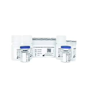
Alpha Complementation Kit Beta-galactosidase
- Original price was: $1,795.00.$895.00Current price is: $895.00.
-
- Sale!
- Research Kits
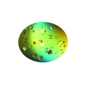
Crystallization Screen for Protein-DNA Complexes
- Original price was: $1,995.00.$995.00Current price is: $995.00.
-
- Sale!
- Molecular Biology, Research Kits
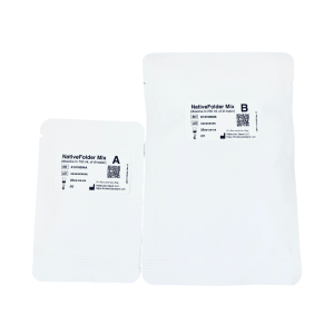
NativeFolder Bacterial Culture Medium
- Original price was: $795.00.$295.00Current price is: $295.00.
-
- Sale!
- Research Kits
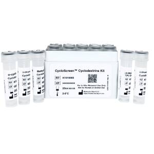
CycloScreen Cyclodextrins Kit
- Original price was: $695.00.$350.00Current price is: $350.00.
-
- Sale!
- Research Kits
NativePro Bradford Protein Assay Kit (Automated)
- Original price was: $995.00.$495.00Current price is: $495.00.
-
- Sale!
- Interference, Research Kits
Rheumatoid Factor Interference Kit
- Original price was: $1,495.00.$695.00Current price is: $695.00.
-
- Sale!
- Interference, Research Kits
Interference Test Kit EXTRA
- Original price was: $1,295.00.$695.00Current price is: $695.00.
-
- Sale!
- Interference, Research Kits
HAMA Interference Kit
- Original price was: $1,295.00.$695.00Current price is: $695.00.
-
- Sale!
- Buffers & Solutions, ELISA, Research Kits
ELISA Microplate Production Kit
- Original price was: $795.00.$495.00Current price is: $495.00.