
A Systematic Review of Sellar and Parasellar Brown Tumors: An Analysis of Clinical, Diagnostic, and Management Profiles.
Authors of this article are:
Alwani MM, Monaco GN, Harmon SM, Nwosu OI, Vortmeyer AO, Payner TD, Ting J.
A summary of the article is shown below:
OBJECTIVE: To systematically review and analyze clinical, diagnostic, and management trends in sellar and parasellar brown tumors reported in existing literature.METHODS: In this systematic review, PubMed, Ovid MEDLINE, Scopus, and Google Scholar databases were searched for reported cases of sellar/parasellar brown tumors. Relevant titles and s were screened in accordance to the Preferred Reporting Items for Systematic Reviews and Meta-Analyses protocol. Articles meeting inclusion criteria were subjected to data extraction, summarization, and analysis. A rare case of parasellar brown tumor was also presented.RESULTS: Eight reports (including the current report) were eligible for inclusion. Mean patient age was 42.75 years. Reported symptoms included visual disturbances (n = 6), headache (n = 5), fatigue (n = 3), nausea/vomiting (n = 2), chest pain (n = 1), neck pain (n = 1), and dysphagia (n = 1). In cases where computed tomography findings were provided (n = 6), lesions were noted to be expansile and lytic. Lesions were hyperintense on T2-weighted magnetic resonance imaging (66.7%) and demonstrated contrast enhancement (83.3%). Histology unanimously showed multinucleated giant cells in a fibrovascular connective tissue stroma. Dramatic symptom resolution was noted in all patients who underwent resection of the sellar/parasellar brown tumor (n = 4; 50%).CONCLUSIONS: Sellar/parasellar brown tumors are a rare, tertiary manifestation of hyperparathyroidism and can be elusive to diagnose. Diagnosis requires a high index of clinical suspicion in addition to comprehensive biochemical testing, imaging, and histopathologic analysis. Surgical extirpation is favored in cases where the lesion is causing compressive symptoms, or if it is unresponsive to management of hyperparathyroidism.Copyright © 2019 Elsevier Inc. All rights reserved.
Check out the article’s website on Pubmed for more information:
[link-preview url=https://www.ncbi.nlm.nih.gov/pubmed/31470158 forceshot=true]
This article is a good source of information and a good way to become familiar with topics such as: Brown tumor; Hyperparathyroidism; Parasellar tumor; Sellar tumor; Sphenoid sinus tumor.
NativeFolder: The Only Bacterial Culture Medium for the Expression of Soluble Proteins
-
- Sale!
- Molecular Biology, Research Kits
NativeFolder Bacterial Culture Medium
- Original price was: $795.00.$295.00Current price is: $295.00.

Interference Test Kit for Assay Validation
-
- Sale!
- Interference, Research Kits
Interference Test Kit
- Original price was: $995.00.$495.00Current price is: $495.00.

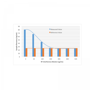
Rheumatoid Factor Interference Blocker
-
- Sale!
- Antibodies, Interference
Rheumatoid Factor Interference Blocker
- Original price was: $595.00.$395.00Current price is: $395.00.

New Antibodies from MOLECULAR DEPOT
-
- Sale!
- Antibodies
Zebrafish wnt8b Antibody (Center)
- Original price was: $1,295.00.$695.00Current price is: $695.00.
-
- Sale!
- Antibodies
TRIB2 Antibody (N-Term)
- Original price was: $1,295.00.$695.00Current price is: $695.00.
-
- Sale!
- Antibodies
NTN1 Antibody (C-Term)
- Original price was: $1,295.00.$695.00Current price is: $695.00.
-
- Sale!
- Antibodies
NME1 Antibody
- Original price was: $1,295.00.$695.00Current price is: $695.00.
-
- Sale!
- Antibodies
Parp9 Antibody (C-Term)
- Original price was: $1,295.00.$695.00Current price is: $695.00.
-
- Sale!
- Antibodies
CDK1 Antibody
- Original price was: $1,295.00.$695.00Current price is: $695.00.
-
- Sale!
- Antibodies
DANRE hoxc11a Antibody (N-Term)
- Original price was: $1,295.00.$695.00Current price is: $695.00.
-
- Sale!
- Antibodies
VDR Antibody (Center)
- Original price was: $1,295.00.$695.00Current price is: $695.00.
-
- Sale!
- Antibodies
ITIH4 Antibody (C-Term)
- Original price was: $1,295.00.$695.00Current price is: $695.00.
-
- Sale!
- Antibodies
SEMA4B Antibody (C-Term)
- Original price was: $1,295.00.$695.00Current price is: $695.00.

New Proteins from MOLECULAR DEPOT
-
- Sale!
- Conjugates, Proteins
Captopril HSA Biotin Conjugate
- Original price was: $1,995.00.$995.00Current price is: $995.00.
-
- Sale!
- Proteins
PKC-α, Active, GST-tagged from Xanopus sp.
- Original price was: $1,595.00.$795.00Current price is: $795.00.
-
- Sale!
- Conjugates, Proteins
Prilocain BSA Biotin Conjugate
- Original price was: $1,995.00.$995.00Current price is: $995.00.
-
- Sale!
- Conjugates, Proteins
T3 (Triiodothyronine) BSA Biotin Conjugate
- Original price was: $1,995.00.$995.00Current price is: $995.00.
-
- Sale!
- Conjugates, Proteins
Pipemidic Acid BSA Biotin Conjugate
- Original price was: $1,995.00.$995.00Current price is: $995.00.
-
- Sale!
- Proteins
PKD2 Protein, Active (Recombinant Human)
- Original price was: $1,795.00.$895.00Current price is: $895.00.
-
- Sale!
- Conjugates, Proteins
Meropenem BSA Biotin Conjugate
- Original price was: $1,995.00.$995.00Current price is: $995.00.
-
- Sale!
- Conjugates, Proteins
Piperacillin BSA Biotin Conjugate
- Original price was: $1,995.00.$995.00Current price is: $995.00.
-
- Sale!
- Proteins
Protein A Peroxidase from Staphylococcus aureus/horseradish
- Original price was: $1,195.00.$595.00Current price is: $595.00.
-
- Sale!
- Conjugates, Proteins
Tobramycin BSA Biotin Conjugate
- Original price was: $1,995.00.$995.00Current price is: $995.00.

New Chemicals from MOLECULAR DEPOT
-
- Sale!
- Chemicals, Interference, Lipids
Triglyceride Mix for Interference Testing
- Original price was: $895.00.$395.00Current price is: $395.00.
-
- Sale!
- Chemicals
Microparticle Stabilizer Solution
- Price range: $295.00 through $495.00
-
- Sale!
- Buffers & Solutions, Chemicals
Enzyme Acceptor Stabilization Buffer
- Original price was: $495.00.$250.00Current price is: $250.00.
-
- Sale!
- Buffers & Solutions, Chemicals
Enzyme Donor Stabilization Buffer
- Original price was: $495.00.$250.00Current price is: $250.00.
-
- Sale!
- Chemicals, Conjugates
Progesterone Biotin Conjugate Solution
- Original price was: $995.00.$595.00Current price is: $595.00.
-
- Sale!
- Buffers & Solutions, Chemicals
Microparticle Activation Buffer
- Price range: $395.00 through $695.00
-
- Sale!
- Buffers & Solutions, Chemicals
Microparticle Washing Buffer
- Price range: $395.00 through $695.00
-
- Sale!
- Buffers & Solutions, Chemicals
Microparticle Blocking Buffer
- Price range: $395.00 through $695.00
-
- Sale!
- Buffers & Solutions, Chemicals
Latex Microparticles Anti Aggregation Agent
- Price range: $295.00 through $3,500.00
-
- Sale!
- Chemicals
CPRG (Chlorophenol red-β-D-galactopyranoside)
- Original price was: $850.00.$395.00Current price is: $395.00.